Agilent BioTek Imaging and Microscopy
Agilent BioTek Lionheart automated microscopes offer powerful microscopy and can easily be equipped with the environmental controls that are crucial for successful short- and long-term kinetic live cell imaging. The Agilent BioTek Cytation C10 confocal imaging reader combines confocal, widefield and multimode detection in a single platform, attainable for every laboratory. The Agilent BioTek Cytation 1, 5, and 7 cell imaging multimode readers offer an array of imaging and multimode capabilities, including digital widefield inverted microscopy, upright microscopy and environmental controls for live cell workflows. Agilent BioTek Gen5 microplate reader and imager software includes functionality for easy image capture and analysis, for both qualitative and quantitative data.
Since its launch, the Cytation line of instruments has continued to evolve, while thousands of researchers around the world have harnessed its capabilities to push the frontiers of scientific knowledge.
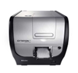 |
Most innovative microplate reader on the market. Added a fully functioning microscope to a plate reader. |
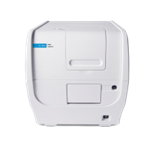 |
Extensively expanded microscopy capabilities to reach a broader microscopy user base while also expanding plate reader capabilities for the drug screening market. |
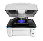 |
Our first automated microscope with an open stage expanding workflow capabilities for live cell imaging market and beyond. |
|
A value-focused Cytation for every lab at a price point that was unheard of for its capabilities. |
 |
Economical automated microscopy at a non-automated microscopy price. |
|
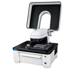
Expanded capabilities for high- speed upright imaging. Applications such as ELISpot and fast slide scanning are now possible. |
 |
The Cytation story is taken to the next level with spinning disc confocal imaging. Enables insights in cutting edge 3D cell models and thick samples. |
![]()
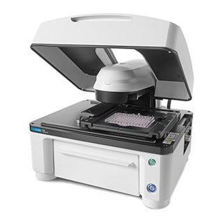
BioTek Cytation C10 confocal imaging reader combines automated confocal and widefield microscopy with conventional multimode microplate reading. The patented confocal design gives exquisite resolution and optical sectioning capabilities for many sample types. A Hamamatsu scientific CMOS camera, Olympus objectives, and laser-based illumination deliver high-quality images.
Cytation C10 also includes widefield fluorescence, brightfield and phase contrast optics. The variable bandwidth monochromator-based multimode plate reading is based on the proven design of the Agilent BioTek Synergy products. Environmental controls, 3D viewer, and Gen5 software enable high-quality results for many applications. The system delivers affordable confocal capabilities to every laboratory.
Lionheart FX automated microscope is a compact system for a broad range of imaging workflows. It offers up to 60x and 100x oil immersion magnification, with fluorescence, brightfield, color brightfield, and phase contrast channels for maximum application reach.
Cytation C10 confocal imaging reader combines automated confocal and widefield microscopy with conventional multimode microplate reading. The patented design gives exquisite resolution and optical sectioning capabilities for many sample types. A Hamamatsu scientific CMOS camera, Olympus objectives, and laser-based illumination deliver high-quality images.

Capture detailed cellular images, get cell-level analyses for migration and wound healing assays, 3D spheroid, and many other kinetic applications. Built-in scheduling, environmental monitoring, and available liquid handling for walk away automation. Multiple users can run simultaneous processes for maximum efficiency in the lab.
"BioTek Lionheart™ FX Automated Microscope is perfect for live-cell imaging. We have been using Lionheart™ FX Automated Microscope for several years. It is an extremely robust system which can handle different tasks. Excellent system for small-medium scale screening. Integrated incubator allows live-cell imaging for prolonged periods. Sensitivity is also excellent."
| Cytation C10 |
Cytation 7 |
Cytation 5 |
Cytation 1 |
Lionheart FX |
Lionheart LX |
|
| Widefield fluorescence | ✔ | ✔ | ✔ | ✔ | ✔ | ✔ |
| Spinning disk confocal fluorescence | ✔ | |||||
| High-contrast brightfield | ✔ | ✔ | ✔ | ✔ | ✔ | ✔ |
| Brightfield | ✔ | ✔ | ✔ | ✔ | ✔ | |
| Color brightfield | ✔ | ✔ | ✔ | ✔ | ✔ | |
| Phase contrast | ✔ | ✔ | ✔ | ✔ | ||
| Live cell incubation option | ✔ | ✔ | ✔ | ✔ | ✔ | |
| Automated plate loading option | ✔ | ✔ | ✔ | ✔ | ||
| Multimode plate reader option | ✔ | ✔ | ✔ | ✔ | ||
| Magnifications available - air | 1.25x to 60x | 1.25x to 60x | 1.25x to 60x | 1.25x to 60x | 1.25x to 60x | 1.25x to 60x |
| Magnifications available - oil | 60x and 100x | 60x and 100x | ||||
| Inverted microscope | ✔ | ✔ | ✔ | ✔ | ✔ | ✔ |
| Upright microscope | ✔ |
Agilent BioTek instrumentation and software create unique augmented microscopy experience with the integration and automation of all steps from image capture to the creation of publication-ready data. There is no need for other software as Agilent BioTek Gen5 does it all quickly and simply:
Augmented Microscopy creates a 'map' with key analytical information, helping you to make informed decisions faster.
The critical first step in a typical microscopy workflow is the efficient and precise acquisition of publication quality images. Augmented Microscopy automates image capture for samples with powerful tools for endpoint and time lapse workflows.

Image processing is essential for optimizing images prior to analysis. Processing tools in Gen5 provide exceptional processing capability to facilitate the analysis of complex biologies.
Captured, processed images are ready for analysis. Image analysis tools in Gen5 cover a very broad range of application requirements, and are both powerful and easy to use. Analysis functions in Gen5 extend to both qualitative and quantitative data.
Augmented Microscopy tools include the ability to create publication-ready images, graphs and data using the functions in Gen5 software. There is no need to export images or data to external software.
Agilent BioTek imaging and microscopy instruments, along with Gen5 software, are capable of automating a broad range of application workflows. Augmented Microscopy tools guide users through the four major steps of microscopy: capture/process/analyze/publish across a broad range of applications. Below are just a few examples of important applications easily managed with Agilent BioTek imagers and Gen5 software.
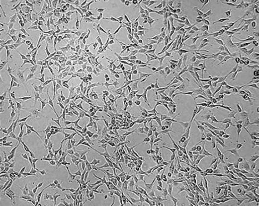
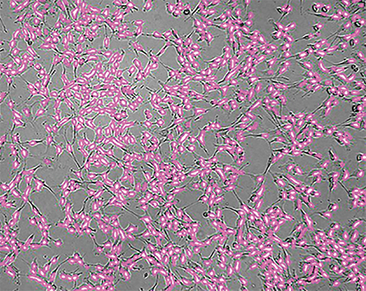
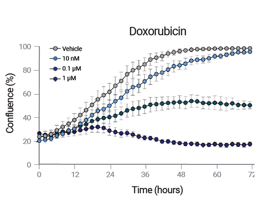
Cells are seeded into 96-well microplates at 2000 cells per well. Environmental conditions, including temperature (37 °C), gas (5% CO2) and humidity (90%) are maintained during a five day incubation by an Agilent BioTek BioSpa 8 Automated Incubator. Proliferation or drug-induced reduction in proliferation is detected by label-free cell counting using high contrast brightfield.
The ELISpot assay monitors ex-vivo cellular immune responses to antigenic stimuli. The two-color format uses two capture antibodies and two independent color developers to quantify the number of PBMCs secreting two separate cytokines. The Cytation 7’s upright color camera is capable of rapidly capturing whole well images for analysis.
Fluorescence immunohistochemistry is a primary technique used in zebrafish imaging research workflows. Optical sectioning is essential for high-quality imaging of deep tissue structures in whole-mount zebrafish, such as the retina and lens of the eye. Spinning-disk confocal microscopy overcomes imaging limitations of widefield microscopy for zebrafish whole-mount immunofluorescence.
RelA, also known as p65, is involved in NF-κB heterodimer formation. With receptor mediated stimulation, NF-κB heterodimers translocate to the nucleus, where it is activated and serves as part of the NF-κB transcription factor complex.
The family of GFP photoproteins are useful tools for live cell analysis. The BacMam baculovirus is an easy to use vector to transiently transfect cells with GFP. The efficiency of the transfection can be determined by counting cells expressing green fluorescence relative to the total number of cells.