Agilent | Trusted AnswersAgilent | Trusted Answers

Complete Your Cell-Based Assay Workflow

With Agilent Microplates

Page Title | Agilent

Use promo code 2257 for a 20% discount!

Sourcing your microplates shouldn’t be an added task in preparing your cell-based workflows. Agilent now offers high quality cell culture and imaging microplates so you can feel confident about your integrated applications, and generate optimal images and results.

Agilent Cell Culture and Imaging microplates offer 96- and 384-well, black and clear wall configurations to meet your fluorescence and label-free assay needs.

Each plate type can simplify your assay procedure and deliver exceptional results across multiple assay workflows, such as:

Reader and Imaging-based Fluorescence Assays

Microplater reader-based fluorescence detection assays can be performed with analytes in suspension, or using cells that are typically adhered to the bottom of the well. Many times it is optimal to perform the plate read from below the plate, as the PMT is closest to the cell layer. 

Fluorescence microscopy includes a variety of assay types, including immunofluorescent staining, as well as assays performed with fluorescent stains or proteins.
The three application notes below demonstrate how Agilent's cell culture and imaging plates can be used for a diverse set of imaging applications.

Automated Cell Fixing and Antibody Staining in Microplates (agilent.com)

High-Throughput Methods to Quantitatively Evaluate TGF-β Signaling in Angiogenesis (agilent.com)


Label-free imaging

Label-free imaging enables researchers to view changes to cell number, as well as morphology, without the use of a label, which may negatively affect cell health.

Read the application note, below, to see how label-free phase contrast imaging is used to aid in proper analysis of single cell object tracking.

Automated High-Throughput Imaging and Analysis of Cell Migration (agilent.com) 


Kinetic live cell assays

Live cell imaging assays, performed over time, enable researchers to gain additional information about cellular responses to treatment. 

The application notes below demonstrate how different kinetic live cell imaging applications can be performed using Agilent imaging microplates.


Automation

Live cell imaging assays, performed over time, enable researchers to gain additional information about cellular responses to treatment.

The ability to incorporate automation into an assay procedure can allow a greater number of samples to be processed per run, increasing assay throughput. By incorporating labware that adheres to ANSI/SLAS standards, one can be confident that each plate or reservoir will be amenable to the automated system of choice.

The application note, below, illustrates how a washer/liquid handler can be used with Agilent imaging plates to perform automated fixing and staining for immunofluorescent assays.


What microplate do you need for these assays?

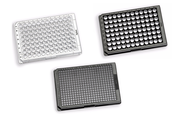
Agilent's cell culture and imaging microplates provide multiple highly sought after features when looking for a high quality imaging plate, including:

 - 96- and 384-well configurations

 - Flat bottom wells

 - Black wall plates contain coverslip (190 µm) thin optically clear well bottom

 All plate definitions are also pre-defined in Bravo and BioTek Gen5 software to ensure the best quality and ease-of-use can be achieved for all cellular imaging applications. 

Order yours today