


Thank you to those who stopped by to see Agilent at SLAS2025! We hope you enjoyed the conference as much as we did. If you missed us, be sure to check out how Agilent can help support your automation needs and bring your lab into the future. Connecting automated solutions to your current workflow allows for current manual processes to be a thing of the past, making your day easier and giving you back more time to focus on other tasks. Learn about the many exciting ways Agilent can help connect you with automation solutions with confidence.
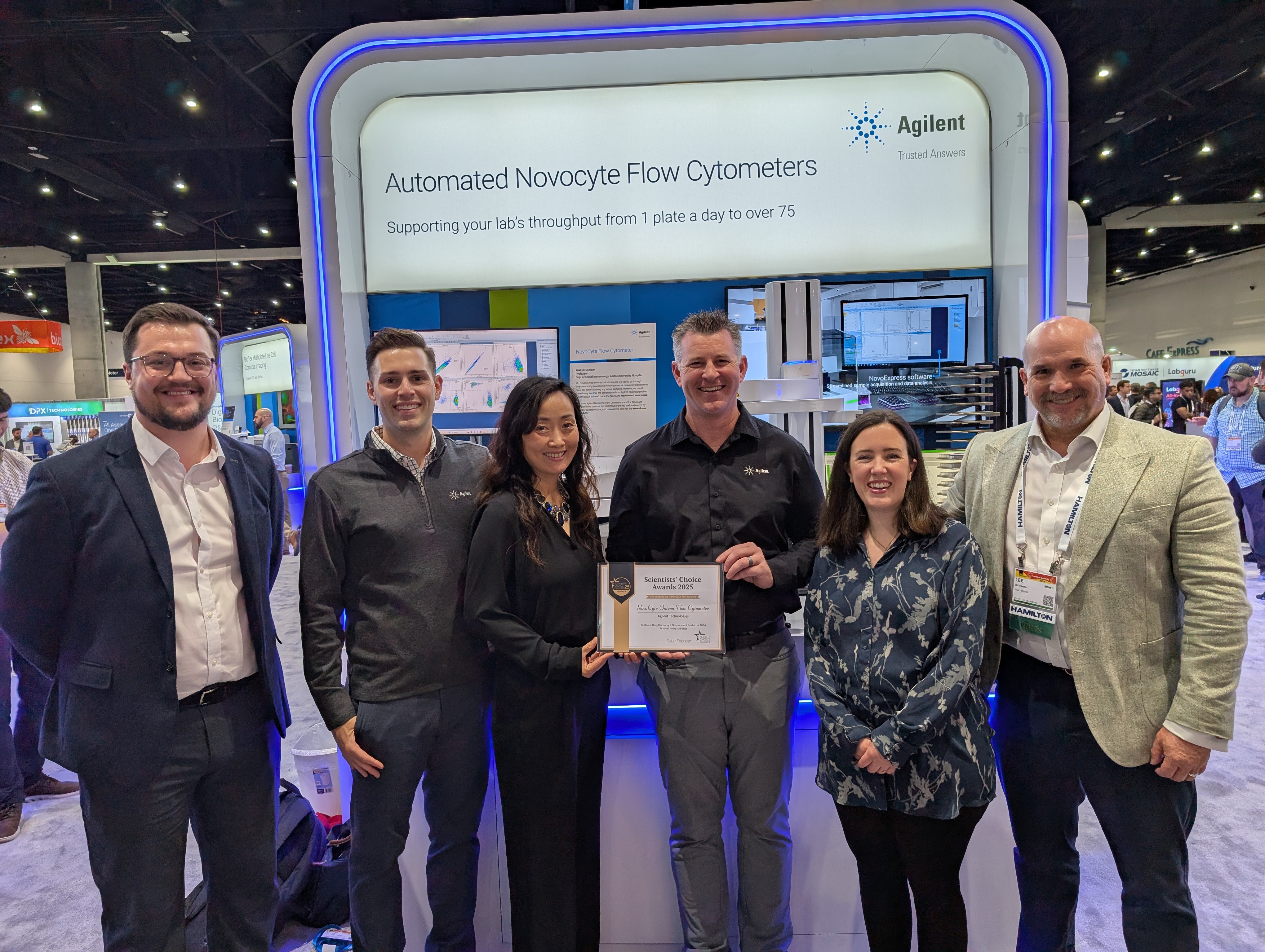
Agilent is the winner of SelectScience's "Scientists' Choice Award" for Best New Drug Discovery & Development Product of 2024 for our Opteon NovoCyte Spectral Flow Cytometer!
Introduction: Due to the diversity and complexity of human rhinovirus (HRV) serotypes, the development of effective vaccines and antiviral therapies remains a challenge. Current viral detection and infectivity assays, such as TCID50 and plaque assays, require prolonged incubation periods and labeling infected cells or counting plaques. Alternative methods introduce additional sample preparation including the generation of genetically modified viruses, or reliance on antibodies. ViQi Inc. has addressed the field's current limitations by developing AVIA (Automated Viral Infectivity Assay) as an efficient solution for viral detection. AVIA’s ability to rapidly detect subtle phenotypic changes in cells post viral infection relies on proprietary state-of-the-art machine learning algorithms to process brightfield images. By detecting the phenotype of the initial cycle of viral infection, AVIA eliminates the need for long incubation times and extensive sample preparation required by traditional assays. Pairing automated image collection on the Agilent BioTek Cytation cell imaging multimode reader with AVIA analysis provides a significantly faster and more reproducible assay for virus titer quantification. Here we showcase an example of AVIA’s utility in quantifying infectivity and experimental monoclonal antibody neutralization results in Vero cells treated with human rhinovirus.
Methods: Vero cells were infected with human rhinovirus (HRV16) and imaged 16 hours post infection using label-free brightfield microscopy at 20x magnification with the Agilent BioTek Cytation 7 cell imaging multimode reader. The ViQi cloud performed a one-time AI training using a single training plate with cells infected at various virus dilutions. Once trained, the AI analyzed assay plates containing multiple dilutions and replicates of the tested infection conditions. Computer vision allows AI infectivity predictions (MOIs, IFU/mL) to be quantified during an initial infection based on phenotypic changes. AVIA employs classification models (convolutional neural networks) to extract image features at increasing levels of abstraction and combines them to make a binary prediction of infection on thousands of sub-image regions. Consequently, unlike the infected/uninfected score in a well of a TCID50 assay, the per-well AIVA infectivity estimate is a continuous value similar to a plaque count. Unlike plaque assays, AVIA does not depend on cell death, allowing for quantification of viruses that do not form plaques.
Results: AVIA identified rhinovirus infection in Vero cells only 16 hours post infection using label-free images of live cells. Automated brightfield images captured on the Agilent BioTek Cytation cell imaging multimode reader supported rapid, high-throughput imaging for downstream analysis. The AVIA approach for viral quantification achieved precision and reproducibility within 10% and detection six times faster than comparative results with a traditional plaque assay.
Acknowledgements: We would like to thank Dr. Tong-Ming Fu and Dr. Yongjun Yu for their important contributions to this work.
For Research Use Only. Not for use in diagnostic procedures.