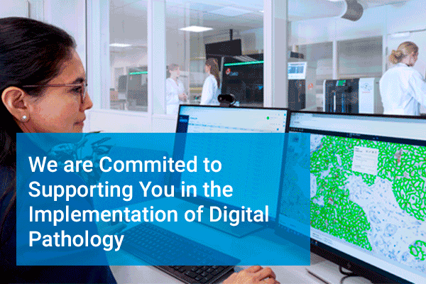
Join our Lunch Digital Pathology Symposium
Agilent will offer a lunch box during the symposium, and you will receive a digital certification of attendance.
Date: Tuesday, 10 September 2024
Time: 13:00-14:30
Location: Room Spadolini 7
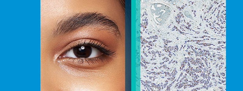
Digital pathology is revolutionizing laboratory efficiency, clinical utility, and quality control, leading to reduced time to diagnosis and improved patient outcomes. This seminar will showcase real-world examples and insights on how scalable, agnostic, end-to-end digital pathology solutions can be effectively utilized to advance pathology labs in practice.
Join us for a lunchtime seminar brought to you by Agilent and in association with digital pathology partners, Hamamatsu, Proscia, Visiopharm and PathAI.
|
|
|
|
|
Biography: I am Professor of Gastro-intestinal and Liver Pathology at Imperial College and Clinical Lead for Gastro-intestinal Pathology in the North-West London Pathology Trust. I also work at the Ludwig Institute for Cancer Research in the University of Oxford. My interests are in clinical and experimental aspects hepato-biliary and gastro-intestinal disease. I am, especially, focused on liver cancer, steatohepatitis and gastro-intestinal malignancy. I am particularly interested in developing new approaches to obtaining information from tissue using computational pathology, metabonomic and other imaging techniques. Abstract: The digitisation of pathology promises to deliver improved workflows and allow for the application to Artificial Intelligence (AI) to challenging areas. Medical liver biopsies are an exemplar of this. Although these biopsies are relatively low volume, they require a high level of specialist reporting of a kind available only in regional centres. Digitisation expedites the transfer of biopsies to these centres and allows for clinical decisions to be made in a time frame that improves patient outcome. We have experience in setting up such a network in NW London. The commonest indication for medical liver biopsy is metabolic dysfunction associated liver disease (MALD). Patients with this condition are increasingly being recruited into clinical trials. Entry is based on semi-quantitative assessment of key histological features. Unfortunately, this assessment has been shown to be very subjective and AI based approaches, in which we are actively involved, have been applied to address the problems posed by MALD. |
|
|
Biography: Teemu Tolonen graduated from the University of Tampere with a Doctorate of Medicine and Doctorate of Philosophy with a focus in cancer genetics. He became an adjunct professor of pathology in 2019. In 2010 he began his residency at Fimlab Laboratories, one of the leading laboratory companies in Finland, providing laboratory services, education, and research in Pirkanmaa, Central Finland, Kanta-Häme, Ostrobothnia and Paijät-Hame regions. In 2017 Dr. Tolonen became the Head of the department of pathology at Fimlab Laboratories. Over the course of his career, Dr. Tolonen has served key roles for the Finnish Division of the International Academy of Pathology (IAP), as a board member (2014-2015) and President (2016-2017/2018-2019). Dr. Tolonen has 62 publications that include scientific publications on genitourinary lab cancers and digital image analysis. Abstract: The integration of digital pathology image management systems (IMS) with embedded multi-AI workflows represents a transformative advancement in the field of histopathology. This presentation will explore the significant value these technologies bring to clinical practice, using IHC quantification as an example, and when deployed at scale in a full software-as-a-service (SaaS) solution across multiple sites and with multiple applications. The session will focus on practical considerations for system requirements, implementation, and future direction of a true multi-site, multi-AI workflow in pathology. |
|
|
Biography: Dr. Bert van der Vegt has been working as a consultant breast and head & neck pathologist at the department of Pathology and Medical Biology of the University Medical Center Groningen, The Netherlands since 2012. His research focuses on the discovery, validation and clinical implementation of biomarkers in cancer (breast and head & neck cancer) and auto-immune disorders (Sjögren’s syndrome). In particular the use of digital- and computational pathology (DCP) in this field has his interest. He is leading the DCP unit of the department of Pathology, which focuses on bringing Digital Image Analysis algorithms to routine clinical pathology practice. Abstract: To really benefit from the advantages of computational pathology these techniques need to be seamlessly integrated in the routine digital pathology practice. This talk will focus on current challenges and possible solutions and opportunities in optimizing the digital pathology workflow from the perspective of a pathology department that has been working fully digital for over 5 years. |
|
|
Biography: Dr. Holger Moch is a Professor of Pathology at the University Zurich, Switzerland. He is the Chairman of the Institute for Surgical Pathology as well as Clinical Co‐Director in the Department Pathology of the University Hospital Zurich. Dr. Moch is a graduate of the Humboldt University Berlin (Charité), Germany and completed his residency training at the Institute for Pathology, University Basel, Switzerland. Dr Moch is a member of the United States and Canadian Academy of Pathology, the editorial board of various journals and boards of several cancer research foundations. He is member of the Executive Committee of the Swiss Group for Clinical Cancer Research (SAKK), the Cancer Network Zurich (CNZ), and the Competence Center for Systems Physiology and Metabolic Diseases (CC‐SPMD) of the University Zurich and the Swiss Federal Institute for Technology Zurich (ETH). His work is described in more than 200 per reviewed papers. In 2007, he was elected as a member of the German National Academy of Science Leopoldina. Evolving Role of Digital Pathology in Anatomic and Molecular Pathology Laboratories |
D0116979_1.00
Please fill out all details required.
 Jake Eden
 Robert D Goldin
 Teemu Tolonen, MD, PhD
 Bert van der Vegt, MD, PhD
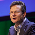 Holger Moch, Professor, MD